The biceps muscle is situated at the front of your upper arm and is responsible for bending your elbow and also rotating your forearm so your palm faces up. One end of the tendon attaches to your ‘radius bone’ in your forearm and the other ends attach up at your shoulder. If you tear or rupture that tendon you will usually have pain and bruising around your elbow and forearm and you may have even heard a pop at the time of injury.
Injury to the Distal Biceps Tendon:
A partial tear means that part of the tendon is still attached. The torn part will not heal back to the bone without surgery. You may be left with persistent pain and weakness when bending your elbow or rotating your forearm.
A complete tear or rupture means that no tendon is left on the bone. You will have weakness which continues long term as well as cramping and early fatigue of that muscle. It will not heal back to the bone without surgery.
Causes and Risk Factors:
A rupture will usually occur if a heavy item is suddenly lifted or a sudden movement of the elbow occurs with a large opposing force.
Risk factors include: steroid use, smoking and advanced age.
What to do if you suspect a torn Distal Biceps Tendon:
If you think you have injured your distal biceps tendon then it is advised to see your GP or Family Doctor for an examination and an Ultrasound request to be obtained. If a rupture or tear is confirmed on Ultrasound then you will be referred to Dr Chiri for an examination and an MRI scan to confirm the extent of the rupture and how far the tendon has moved away from the bone.
Treatment:
During surgery there is a cut at the front of your forearm and sometimes extended in to the elbow if the tear has been there for a long time. The end of the tendon is identified and prepared for repair.
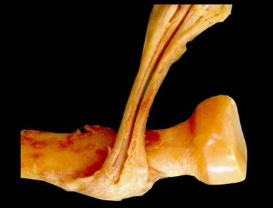
Dr Wael Chiri performs a technique which repairs the distal biceps tendon back to where it originally was. This is a technique which was published by Professor Greg Bain (Adelaide, South Australia). Classically the tendon is repaired close to its original position as seen below.
Credit to: Matthews, E. C., et al. "The biotenodesis screw and endobutton technique for repair for acute distal biceps tendon rupture." Clin Arch Bone Joint Dis 1.006 (2018).
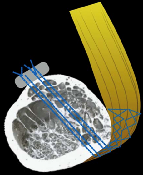
However the tendon does not attach to that part of the bone as seen on images below. It attaches to the side of the bone rather than directly on top.
Credit to: Professor Greg Bain. Accessed via Phadnis, J. and Bain, G., 2015. Endoscopic-assisted distal biceps footprint repair. Techniques in Hand & Upper Extremity Surgery, 19(2), pp.55-59.
Therefore in the ‘anatomic repair’ method the button sits on the top of the bone and the tendon heals on the side of the bone where it belongs.
Professor Greg Bain. Accessed via Phadnis, J. and Bain, G., 2015. Endoscopic-assisted distal biceps footprint repair. Techniques in Hand & Upper Extremity Surgery, 19(2), pp.55-59.
Rehabilitation:
The patient leaves hospital in a sling and wears that sling for comfort. They are able to do range of motion but strictly no moderate/heavy lifting for 3 months.